샘창자걸이근
package.lua 80번째 줄에서 Lua 오류: module 'Module:Namespace detect/data' not found.
| 샘창자걸이근 | |
|---|---|
 | |
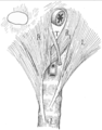
| 샘창자의 그림. 샘창자걸이근은 그림에서 보이는 샘빈창자굽이(duodenojejunal flexure)에 붙는다. | |
 | |
| 샘창자걸이근은 그림에 나타난 이자의 뒤에서 샘빈창자굽이 뒤쪽으로 닿는다. | |
| 영어 명칭 | suspensory muscle of duodenum |
| 기관 | 위장관계 |
| 신경 | 복강신경얼기, 위창자간막신경얼기 |
샘창자걸이근(suspensory muscle of duodenum) 또는 십이지장제근(十二指腸提筋)은 샘창자와 빈창자 사이의 접합부인 샘빈창자굽이를 복강동맥과 위창자간막동맥을 둘러싸는 결합조직으로 연결하는 얇은 근육이다. 트라이츠 인대(ligament of Treitz)로도 알려져 있다.[1] 샘창자걸이근은 대부분 샘창자의 셋째, 넷째 부분과 샘빈창자굽이에 모두 연결되어 있지만 이 붙는 곳은 변이가 크다.
샘창자걸이근은 marks the formal division between the first and second parts of the small intestine, the duodenum and the jejunum. This division is used to mark the difference between the upper and lower gastrointestinal tracts, which is relevant in clinical medicine as it may determine the source of bleeding in the gastrointestinal tract.
The suspensory muscle is derived from mesoderm and plays a role in the embryological rotation of the gut, by offering a point of fixation for the rotating gut. It is also thought to help digestion by widening the angle of the duodenojejunal flexure. Superior mesenteric artery syndrome is a rare abnormality caused by a congenitally short suspensory muscle.
구조[편집]
샘창자와 빈창자는 각각 작은창자의 첫 번째, 두 번째 부분이다. 샘창자걸이근은 이 두 부분을 나누는 경계이다.[2] 샘창자걸이근은 가로막의 오른쪽 다리에서 일어나 식도 주변을 주행한다. 이후 복강동맥 줄기와 위창자간막동맥 주변의 결합조직으로 연속되어 이자 뒤쪽을 지나고, 창자간막의 위쪽 부분으로 들어가 샘창자와 빈창자 사이의 접합부인 샘빈창자굽이에 닿는다.[3] 여기서 샘창자걸이근은 생샘창자의 근육층으로 연속된다.[1]
변이[편집]
길이와 근육이 붙는 곳에 대해서 고려할 만한 해부학적 변이가 존재한다.[4] 고전적인 샘창자걸이근에 대한 설명과 다르게, 이 근육이 샘빈창자굽이에만 닿는 경우는 전체의 8% 정도이다. 훨씬 더 많은 수인 40-60%에서는 샘빈창자굽이에 더해 샘창자의 셋째, 넷째 부분에도 닿으며 20%의 사람들에서는 아예 셋째, 넷째 부분에만 닿는다. 또한 서로 분리된 여러 부분에 붙는 경우도 그리 드물지 않다.[1]
According to some authors, who use the original description by Treitz, the muscle may be divided into two sections: a ligamentous portion attaching the right crus of diaphragm to the connective tissue surrounding the coeliac artery and superior mesenteric artery; and a lower muscular portion from the connective tissue attaching to the duodenum. The superior portion is also described as the Hilfsmuskel.[3][4] These two parts are now considered anatomically distinct, with the suspensory muscle referring solely to the lower structure attaching at the duodenum.[1][4]
Function[편집]
The ligament contains a slender band of skeletal muscle from the diaphragm and a fibromuscular band of smooth muscle from the horizontal and ascending parts of the duodenum. When it contracts, by virtue of connections to the third and fourth parts of the duodenum, the suspensory muscle of the duodenum widens the angle of the duodenojejunal flexure, allowing movement of the intestinal contents.[1][5]
Embryology[편집]
Embryologically, the suspensory muscle of the duodenum is derived from mesoderm. It plays an important role in the embryological rotation of the small intestine as the superior retention band.[1][3]
Clinical significance[편집]
This ligament is an important anatomical landmark of the duodenojejunal flexure, separating the upper and lower gastrointestinal tracts. For example, bloody vomit or melena, black tarry stools, usually indicate a gastrointestinal bleed from a location in the upper gastrointestinal tract. In contrast, hematochezia, bright red blood or clots in the stool, usually indicates gastrointestinal bleeding from the lower part of the gastrointestinal tract.[6] It is an especially important landmark to note when looking at the bowel for the presence of malrotation of the gut, a syndrome often suspected in young children when they have episodes of recurrent vomiting. Visualising a normal location of the ligament of Treitz in radiological images is critical in ruling out malrotation of the gut in a child; it is abnormally located when malrotation is present.[4]
During a Whipple's procedure, commonly used to treat pancreatic cancer by removing the pancreas, duodenum, and part of the jejunum, the ligament of Treitz is separated from the duodenum and preserved. When the remaining jejunum is anastamosed with the pylorus of the stomach, it may be passed through the ligament.[7]
Superior mesenteric artery syndrome (SMA) is an extremely rare life-threatening condition that can either be congenital and chronic, or induced and acute. SMA Syndrome is characterised by compression of the duodenum between the abdominal aorta and the superior mesenteric artery, and may—when congenital—result from a short suspensory muscle. One surgical treatment is Strong's operation, which involves cutting the suspensory muscle, though this is not often carried out.[8]
History[편집]
The suspensory muscle of the duodenum was first named in 1853 by Václav Treitz, as the musculus suspensorius duodeni (in Latin), and described as consisting of a lower muscular portion with a broad base, and an upper tendinous portion blending with connective tissue around the origins of the superior mesenteric and coeliac arteries. It is commonly termed the ligament of Treitz by clinicians and as the suspensory muscle of the duodenum by anatomists. It has also been likened to "a polar ice cap ... a structure that many refer to but few have seen."[1]
추가 이미지[편집]
-
샘창자걸이근의 이는곳인 오른쪽 가로막다리의 섬유가 그려져 있다.
-

샘창자걸이근을 배쪽에서 본 그림.


참고 문헌[편집]
- ↑ 1.0 1.1 1.2 1.3 1.4 1.5 1.6 스크립트 오류: "Citation/CS1" 모듈이 없습니다.
- ↑ 스크립트 오류: "citation/CS1" 모듈이 없습니다.
- ↑ 3.0 3.1 3.2 스크립트 오류: "citation/CS1" 모듈이 없습니다.
- ↑ 4.0 4.1 4.2 4.3 스크립트 오류: "Citation/CS1" 모듈이 없습니다.
- ↑ 스크립트 오류: "citation/CS1" 모듈이 없습니다.
- ↑ 스크립트 오류: "Citation/CS1" 모듈이 없습니다.
- ↑ 스크립트 오류: "Citation/CS1" 모듈이 없습니다.
- ↑ 스크립트 오류: "Citation/CS1" 모듈이 없습니다.
모듈:Authority_control 1173번째 줄에서 Lua 오류: attempt to index field 'wikibase' (a nil value).
This article "샘창자걸이근" is from Wikipedia. The list of its authors can be seen in its historical and/or the page Edithistory:샘창자걸이근. Articles copied from Draft Namespace on Wikipedia could be seen on the Draft Namespace of Wikipedia and not main one.

|
This page exists already on Wikipedia. |